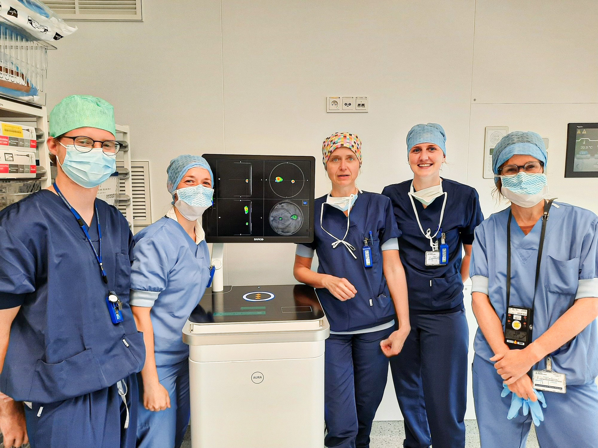
First in our hospital: use of mobile PET-CT scanner on a thyroid cancer patient
Great news from Maria Middelares General Hospital' operating facilities: the first operation using a mobile PET-CT scannertook place in June. The device scans the excised tissue sample and verifies whether any tumour tissue is present (completely) in the sample. Within ten minutes, the surgeon receives a detailed image of the excised tissue so that, even during the procedure, the care professionals can determine whether the tissue is complete.
In a world first, our hospital used the scanner for the first time on a thyroid cancer patient. Dr Valérie Vergucht of the surgery department: ‘We’re proud that this approach allows us to acquire more information during surgeries and hope, that this means operations will have a greater chance of achieving their goal. My thanks to the nuclear medicine and general surgery departments, among others, who have started working with the mobile PET-CT scanner.’
The device used is the AURA 10 PET-CT, developed by the Ghent-based startup XEOS. After the summer, a broader study involving different disciplines will start at Maria Middelares General Hospital to further evaluate the use of this technique.

Something wrong or unclear on this page? Report it.


