Chest X-ray
What is it?
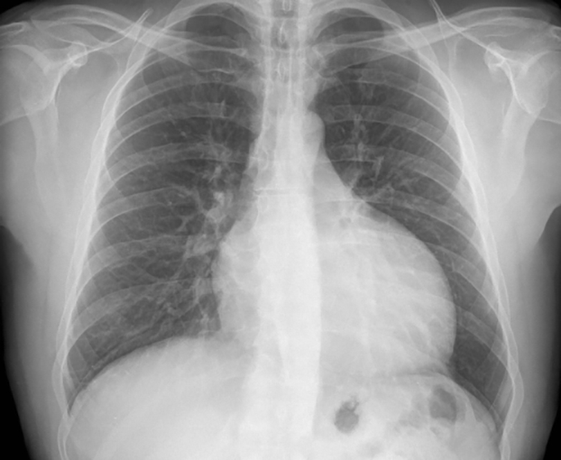
What is it?A chest X-ray is an X-ray photo of the chest, the lungs, the aorta, the heart, the vertebral column and the ribs.
What is the process?
What is the process?You will be placed between a plate that is sensitive to X-rays and an X-ray machine. After inhaling deeply, the image will be taken from an anterior-posterior view and from the side.
What are the risks?
What are the risks?No side effects are expected as the radiation dose is low.
Results
ResultsMedical imaging of the organs inside the rib case help determine a diagnosis. A chest X-ray can help detect:
- an enlarged heart
- fluid accumulation in or around the lungs
- widened aorta
- pulmonary infection
- pneumothorax
In addition, a chest X-ray can help evaluate the position of the electrodes (pacemaker or defibrillator).
Centres and specialist areas
Centres and specialist areas
Heart Centre
A dynamic team with expertise in treating heart problems

Cardiology
Heart and vascular disease
Latest publication date: 16/05/2024
Supervising author: Dr Provenier Frank