Contrast echocardiography (heart)
What is it?
What is it?During echocardiography, contrast medium is sometimes injected through an IV into a vein in the arm. This is called contrast echocardiography.
Contrast medium allows for the detection of certain heart abnormalities.
Preparation
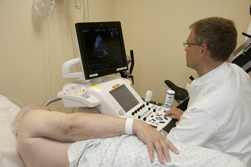
PreparationThree electrodes on the chest are used to monitor the heart rhythm. To get a good view of the heart, you will be asked to lie on your left side.
The treatment room is darkened to allow the cardiologist to have the best view of echocardiography image.
To administer the contrast medium, an infusion is placed in the arm.
Before administering the contrast medium, the physician will check that you do not have any contraindications that would prevent us from administering it to you.
Test
TestAn echocardiography uses a special instrument called an echo probe. This is a type of sensor that emits and captures sound waves. To enable proper conduction, a cold gel is applied to the chest first.
The echo probe is then placed on the chest wall at different points to view the heart from different angles. Sometimes you will be asked to hold your breath for a few seconds during the examination.
It is important to note that the examination is not painful. You will only feel vibrations caused by the echo probe. The whole examination usually takes no more than 30minutes. It helps physicians assess the structure and function of the heart and detect any abnormalities.
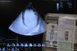
Two types of contrast medium are used.
1. Agitated saline solution.
This is a standard infusion solution (NaCl 0.9%). Of this, 8 cc is mixed with a small amount of air (2 cc). This will cause the cavities of the heart to glow white on the echocardiography screen after injection. This type of contrast medium is used primarily to detect connections between the right and left heart. As this concerns a saline solution mixed thoroughly with a very small amount of air, this technique is completely safe.
2. Special echocardiography contrast fluid: Sonovue
This is a commercially available contrast fluid that fills up the heart cavities, which allows for better assessment of wall motion. In certain circumstances, this product should not be used. So your physician will always study your records carefully before administering this product.
Results
ResultsAt the end of the consultation, the cardiologist will discuss the findings and possible changes in treatment with you.
Aftercare
AftercareAfter the test, the IV will be removed and an adhesive bandage will be placed over the injection site.
No specific aftercare is required.
Centres and specialist areas
Centres and specialist areas
Latest publication date: 16/05/2024
Supervising author: Dr Provenier Frank